This is a long page and will link to various pages that cover the deep dives into different structures and systems.
The regions covered are:
- The skeletal structure of the cranium
- Form and major divisions of the cranium
- Anatomy of the meninges
- Blood supply and venous drainage of the brain
- Ventricular system
- Brainstem and cerebellum
- Cranial nerves
- Forebrain features and functions of each lobe
- Motor and sensory pathways
- Visual and auditory pathways
- Hippocampus
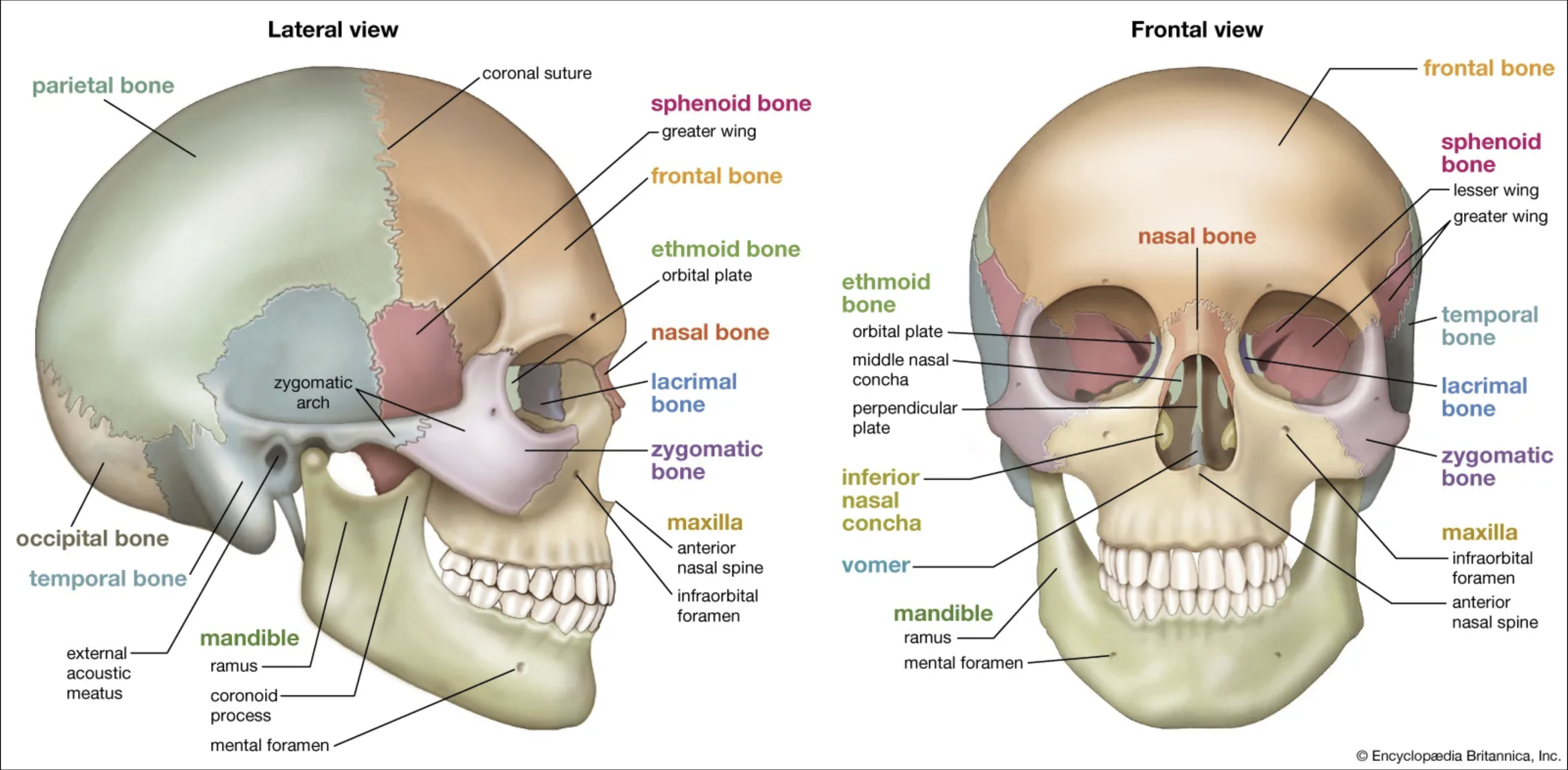
The skeletal structure of the cranium
The Skeletal structure of the cranium is somewhat simple, with 8 bones that make up the cranium. This is relates back to respiratory in MED2 and is continued more.
The cranium is the “container” which holds the brain and is made of 8 bones:
- Frontal bone
- 2x Parietal bones
- 2x Temporal bones
- Occipital bone
- Ethmoid bone
- Sphenoid bone
These are arranged as such:
The cranial flat bones join with sutures, which are the coronal, sagittal, lamboid, and squamous. the t intersection where the coronal and sagittal suture join is called the bregma, and the t intersection where the sagittal and lambdoid sutures join is called the lambda.
In the inferior region of the internal skull there are 3 pairs of “pools” called fossa, where the frontal lobes, temporal lobes and cerebullum sit. They are easily named the anterior middle and posterior fossa. There are many structures in the medial portion of the cranial floor where there are holes for entry and exit for cranial nerves, blood vessels, sinuses and a big one for the spinal cord.
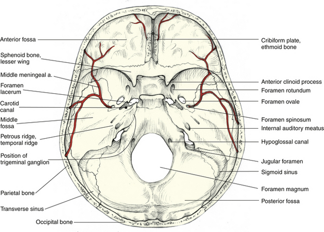 Structures of note in the cranial floor are:
- petrous bone of the temporal bone, extremely strong
- cribriform plate of the ethmoid bone, for the olfactory nerves from the olfactory bulb to pass through to the nasal cavity
- sella turcica of the sphenoid bone, for the pituitary gland to sit inside of
Foramina for the cranial nerves to pass through are listed, and are good to remember:
- Olfactory foramina (cribriform plate), this is for CNI for smell
- Optic foramen, this is for CN II for sight.
- Superior orbital fissure for CN III, IV, V (first branch), VI for oculomotor and a number of other functions. This cannot be seen on the diagram.
- Foramen rotundum for CN V (second branch)
- Foramen ovale for CN V (third branch)
- Internal auditory meatus for CN VII and VIII
- Jugular foramen this is for the CN IX X XI
- Hypoglossal canal which is for the CN XII
Form and major divisions of the Brain.
This is is more of an overview and each section will be expanded on later but its good for an initial view.
Your brain has 3 or so main sections, with each bit subdivided. In general there is the forebrain, midbrain and hindbrain. The forebrain is the cerebral hemispheres and diencephalon, the midbrain is the superior section of the brainstem. The hindbrain is composed of the pons and medulla, the other 2 parts of the brainstem, as well as the cerebellum.
Often people just say cerebrum, brainstem and cerebellum.
Forebrain:
The forebrain is the cerebrum and diencephalon. The cerebrum is responsible for personality, logic, and a lot of higher order thinking. It is split into lobes which have semidistinct functions.
The lobes are:
- Frontal
- Temporal
- Parietal
- Occipital
- and insular
The cerebrum also contains the basal nuclei which are neuron cell bodies that are deep in the cerebrum, not cortical like most of grey matter.
The basal nuclei have 3 regions, the caudate nucleus, the putamen and the globus pallidus.
the lenticular nucleus is a name referring to the putamen and globus pallidus and the striatum is a name referring to the caudate nucleus and the putamen.
The origin of these names comes from the appearance and proximity, which should be easier to understand in the labs.
The function of the basal nuclei are to inhibit unwanted actions, amongst others.
The cerebrum has 2 hemispheres and a structure called the corpus callosum, which is a bundle of commissural fibres which connects the 2 hemispheres.
The diencephalon is a distinct region to the cerebellum and contains the thalamus, hypothalamus epithalamus, and subthalamus. The diencephalon is considered the “gateway” to the cerebrum and all signalling to and from the cerebrum must pass through here.
There is some attention drawn to the arrangement of the diencephalon and basal nuclei
Anatomy of the meninges
This is content we have previously covered but is good to know again.
The meninges are a system of 3 layers which protects, supports, and nourishes the brain as well as allows csf fluid to flow around the brain and be drained.
The three layers each have a specialisation and are detailed below
Dura mater
This is the outermost layer and means tough mother. it has 2 layers, the meningeal and periosteal layers, which diverge at 3 large infoldings which extend down the transverse fissure, inbetween the cerebrum and cerebellum and the 2 hemispheres of the cerebellum.
These are called the falx cerebri, tentorium cerebelli and falx cerebelli. At the base (and inferior edge of the falx cerebri) of each of these foldings there are channels called sinuses and these allow the flow of circulated csf and venous blood ultimately into the internal jugular vein.
The arachnoid mater and pia mater both also fold down into these dural reflections.
Arachnoid mater
This is the middle most layer and its name and primary function is associated with the subarachnoid space, an unfilled areas surrounding the brain that the csf flows around as it passes to the sinuses it also is where blood vessels run as they go around the brain. Its name is from little projections down to the pia mater which have a structural role.
Pia mater
This is the layer closest to the brain and descends down with the sulci.
Blood supply and venous drainage of the brain
Supply
The blood supply to the brain initially comes from the internal carotid arteries, a branch of the common carotid arteries. the ICAs branch into the anterior and middle carotid arteries. The posterior carotid arteries branch from the basilar artery which is a merge of the vertebral arteries. Often there are communicating arteries between these, the anterior communicating artery connecting the anterior cerebral arteries and the posterior communicating artery connecting the internal carotid arteries to the posterior cerebral arteries. This structure which forms the connections between ACA, MCA, and PCA is called the circle of henle.
The anterior cerebral artery is supplies the anterior 2/3rds of the medial are of the cerebrum. The Middle carotid artery supplies the lateral 4/5ths of the cerebrum and the posterior cerebral artery supplies the medial and lateral rear brain (think occipital lobe inferior temporal lobe and superior brainstem.)
the middle meningeal artery is a branch of the external carotid artery and supplies a good portion of the cranial bones and dura mater. it is vulnerable to bleeds.
The inferior brainstem and cerebellum are supplies by the basilar artery and vertebral arteries, with notable offshoots being the superior cerebellar artery, anterior inferior cerebellar arteries (AICA) and the posterior inferior cerebellar arteries (pica)
Drainage
The venous blood of the brain flows into the sinuses of the meninges. and all ultimately flows into the internal carotids.
the superior sinus is where CSF flowing over the lateral brain ends up and the straight sinus is where the medial csf ends up.
The superior sinus and the straight sinus meet at the confluence of sinuses where the transverse sinuses channel it to the sigmoid sinus and to the IJV
The inferior structures drain into the cavernous sinuses where they can flow into the inferior petrosal sinus which flows into the IJV directly or the superior petrosal sinus which flows into the transverse sinus to the sigmoid sinus and IJV.
Ventricular system
The ventricular system has been covered before and so we know there are 4 ventricles, or hollow spaces containing CSF, the liquid that nourishes and feeds the brain cells as well as remove wastes.
The fluid is filtered from blood by the choroid plexi and released to the ventricular system. The lateral ventricles are the first and then the csf moves to the third ventricle, then the cerebral canal and then the fourth ventricle.
From there it flows out of the 2 lateral apertures and median apertures. Then it flows around the brain and spinal cord to the sinuses and are drained.
After flowing out of the apertures it works its way around the outside of the brain, and there are subarachnoid cisterns, which are like spaces that contain relatively more csf, analogous to ventricles but smaller. The names of these are the: Cisterna magna, pontine cistern, interpeduncular cistern, and superior cistern.
choroid plexi
the inferior medullary velum has it but the superior does not.
Brainstem
The brainstem is made of 3 parts: the midbrain, pons and medulla oblongata.
It is a compact and intricate structure with lots of tracts and pathways as well as nuclei for processing. It is also where 10 of the 12 cranial nerves emerge from.
The brainstem is made of 3 sections, the midbrain, pons and medulla.
The midbrain
The midbrain is immediatly below the diencephalon and is a big bridge for motor sensory and autonomic function (basically everything).
Important superficial structures are:
- Cerebral peduncles Crus Cerebri - little feet of cerebrum NOT cerebellar peduncles these are fibres descending from the cores to the brainstem and spinal cord. They also help anchor the cerebrum to the brainstem.
- Corpora quadrigemia Quadruplets This is a group of 4 colliculi (hills), a pair of superior colliculi which deals with certain visual signals (like fast moving or flying objects) and the inferior colliculi which deals with reflexive responses to noises which startle you.
- Substantia nigra Black substance These are neurons which contain melanin, as it is a chemical precursor to dopamine. They are involves with dopamine release and are thus atrophied in parkinsons disease. It is functionally linked to the basal nuclei
- Red nucleus This has extensive blood supply, leading to its red colour. it is a relay nucleus in some motor pathways.
- Cerebral aquaduct This is the passage from the 3rd to 4th vetnricle
- superior cerebellar peduncles These are 1 of 3 peduncles and connect the cerebellum to the brainstem and CNS
The pons
The pons means bridge, and it is a major connection between the cerebral cortex, cerebellum and spinal cord. It also had nuclei involved with breathing sleep-wake cycles and arousal. it also the origin of 4 cranial nerves, 5-8.
it had the pontine nuclei and the pneumotaxic centre, together with the medullary centre
The medulla
The medulla connects the pons to the spinal cord and the cerebellum to the spinal cord. It has the pyramidal decussation and 4th ventricle. It also is the origin of cranial nerves 9-12. It has many reflex centres which are important in controlling heart beat, vasocular tone, breathing as well as more refelxes.
Cerebellum
The cerebellum is a structure highly involved at ensuring the movement plans are refined, realistic (eg in line with the current body position), and general coordination.
Voluntary movements can occur without the cerebellum but they are clumsy and disorganised. (drunken gait for example is similar)
Anatomy of the cerebellum
The cerebullum has 2 hemispheres demarcated by the anterior cerebellar incisure, posterior cerebellar incisure, and the vermis. each hemisphere has 2 lobes, the anterior and posterior. However the functional divisions lie across different boundaries.
There are 3 main divisions of the hemispheres,
- Spinocerebellum This is the medial 50% of the cerebellum and works to balance the posture and movement of the trunk and limbs
- Cerebrocerebellum This is the lateral portions of the cerebellum and are important for planning movement
- Vestibulocerebellum The is is the flocculi and the nodulus which do balance, head and eye movements
-
- deep nuclei The deep nuclei are 3 pairs of nuclei, the dentate, interposed and fastigial, which act to do calculations to refine the
Cerebellum physiology
There are 3 peduncles which connect the cerebellum to the brainstem, there is the superior, middle and inferior. These dont really look like distinct structures and are more sections of one large peduncle.
The SCP is most efferent information away from the cerebellum, and the MCP is mainly afferent to the cerebellum. The ICP does both directions.
Inputs: Inputs to the cerebellum are sensory data from the limbs, and motor plans from the motor cortex and premotor cortex.
Outputs: outputs to the spinal cord and the brainstem which directly influence motor control. and outputs tot he motor areas in the thalamus and the brainstem which helps refine movements.
Cerebellar cortex vs DCN
The cerebellar cortex is involved with comparing sensory data to motor data but the movement refinement, on the go correction as well as learning is chiefly handled by the DCN.
Some symptoms of cerebellar dysnfucntions are loss of cooridination in muscle activity which can present as:
- Ataxia Inaccuracy in the speed force and distance of a movement.
- Tremor Involuntary oscillation of the limbs or trunk
- Nystagmus Rhythmic involuntary oscillation of the eyes
- Headache & vomiting
Cerebellar dysfunction
Cranial nerves
| Nerve | Function | Description |
|---|---|---|
| 1. Olfactory nerve | Special sense of smell | Synapses with the olfactory bulb where nerve endings pass through the cribriform plate of ethmoid bone |
| 2. Optic nerve | Special sense of vision | optic canal → optic chiasm → optic tract → some go to superior colliculi some go to the thalamus |
| 3. Oculomotor nerve | Motor to all extraocular muscles except lateral rectus and superior oblique Motor to intrinsic motors of the eye | Midbrain origin. Exits cranium via the superier orbital fissure |
| 4. Trochlear nerve | Motor to superior oblique muscle | Midbrain origin. Only does 1 muscle so small. originates from dorsal surface of brainstem. As it goes to the eye it exits via the superior orbital fissure |
| 5. Trigeminal nerve | Somatic sensation of face and cavities Motor to muscles of mastication | Pons origin. Fat nerve. 3 divisions which exit at different places. Exits at superior orbital fissure, foramen rotundum, foramen ovale. |
| 6. Abducens nerve | Motor to lateral rectus muscle | Pons origin. As it goes to the eye it exits via the superior orbital fissure |
| 7. Facial nerve | Motor to muscles of facial expression Taste on anterior tounge | Pons origin. Exits via the internal auditory meatus & facial canal |
| 8. Vestibulocochlear nerve | Special senses of hearing and balance | Pons origin. Exits via the internal auditory meatus |
| 9. Glossopharangeal nerve | Major nerve for taste Sensation on posterior tongue and pharynx | Medullary origin. Exits via jugular foramen |
| 10. Vagus nerve | motor and sensory to viscera and thorax and abdomen Motor to muscles of Pharynx and larynx | Medullary origin. Exits via jugular foramen |
| 11. Accessory nerve | Motor to sternomastoid and trapezius msucles | Medullary origin. Exits via jugular foramen |
| 12. Hypoglossal nerve | Motor to all muscles of tongue | Medullary origin. Exits via Hypoglossal canal |
Forebrain features and functions of each lobe
The cerebrum has many different areas, with each having a specific area which it does well
- Brocas area, made of opercular and triangular gyri Deals with spoken speech
- Prefrontal cortex This deals with intellect complex learning, cognition, recall anbd personality. it is heavily dependant on social environment. damage can result in mental and personality disorders
- Supramarginal gyrus This deals with tactile sensory data and in involved with spacial perception and limb spacial awareness
- Primary visual cortex This is the gyri forming the walls of the the calcarine sulcus.
- Visual association area
- uncus
- Amygdala
- Wernickes area deals with listening to communications
- primary auditory cortex
- right hippo campus - spatial representation.
- Insular lobe Function is unknown. visceral sensation effected as well as general sensory and mtoro function. A case study apparently found the left insular lobe was involved in the normal emotional processing of music.
- pineal gland
- thalamus
- hypothalamus
Motor and sensory pathways
Motor pathways
The corticospinal tract starts with the Primary motor, premotor and sensory cortices and then the tract follows through the internal capsule, to the midbrain pons and medulla. most of the fibres (90%) decussate in the medulla and travel down the lateral corticospinal tract on other side of the body to the origin (contralateral). The endpoint is the cell bodies in the ventral horn of the spinal cord which then link to the effector.
Role of the cerebellum.
The cerebellum receives inputs form the motor cortex which relays the intended movements as well as inputs from the somatosensory neurons. The DCN will do comparative calculations and sends corrections to the motor neurons as well as brain stem nuclei.
Role of the Basal nuclei in motor control.
The role of the BN are to recieve inputs from all cortical areas and project back to premotor cortex as well as the prefrontal cortex. it is involved in more abstract (complex) parts of motor control for example broadly inhibiting during rest or stillness and then coordinating motions when the motor centers are release from forementioned inhibition.
Somatosensory pathways
There are 2 main types of pathway to the somatosensory cortex.
The discrimative pathway relates fine touch, vibration and conscious proprioception to the SSC with the first neuron being in the spinal root ganglion, second order neuron being in the medulla where decussation happens, and 3rd order being in the thalamus.
The difference is the nondiscriminatory pathway (which relates pain pressure and temperature) has the decussation and second order neuron in the dorsal horn of the spinal cord.
Spinocerebellar tract
This is a tract which does not contribute to somatic sensation but just sends data to the cerebellum for processing there. first order neuron is in the dorsal root ganglion and the second order neuron is in the dorsal horn with NO DECUSSATION. It is an ipsilateral pathway so right arm position sent to right cerebellar cortex
Visual and auditory pathways
Visual pathway
The retinal cells get excited and send information to the optic nerve where a decussation happens at the optic chiasm. Then optic tract travel to the lateral geniculate nucleus of the thalamus where processing occurs. After this visual information is relayed to the visual cortex. The superior colliculus is involved in tracking fst moving object and startling imagery.
the PVC provessing basic visual information, and conscious perception of visual information.
visual association areas process visual information concerned with form colour and movement.
complec visual proccessing occurs in ventral parts of the temporal lobe, parietal cortex and frontal corex.
- ventral temporal lobe identifies what the object is
- parietal lobe assesses where the location is
- frontal cortex uses visual information to guide movements.