The cranial nerves 1-12 are nerves that originate directly from the brainstem (except I and II) and go to structures in the head neck and torso. They all stem from the brainstem.
Nerves vs nuclei
The difference between cranial nerves and brainstem nuclei are that cranial nerves are axon bundles from the nuclei and can possess a number of types of information. Brainstem nuclei are pockets of neuron bodies in the brainstem where each one has a specific type of information and function. each nucleus can only do either
- general motor
- branchial motor
- visceral motor
- somatic sensory
- visceral sensory
- or special sensory.
Multiple nerves can attach to a single nuclei. Multiple nuclei can associate with a single nerve. Sometimes nerve nuclei pairs are 1-1.
John Reynolds neurodevelopmental map of the nuclei
Quick table
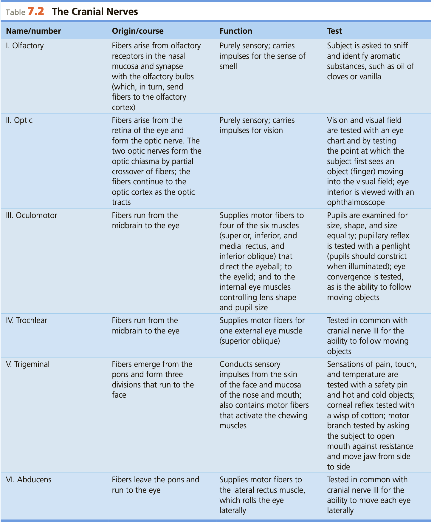
Cranial nerve 1: Olfactory (I)
Function
This is the nerve which enables the special sense of smell.
This is one of the 2 nerves that do not originate from the brainstem, the other being the optic nerve.
Test
The test for this is to have the patient smell something, like vanilla or something.
Origin/pathway
The nasal fibres in the nasal cavity, which then travel up, through the holes in the cribriform plate in the skull to the olfactory bulb, where they synapse and then travel to the primary olfactory cortex of the inferior temporal cortex. As it does not connect to the brainstem it does not have a brainstem nuclei.
Associated cerebral gyrus/area
As this nerve does not join with the brainstem it does not have an associated brainstem nuclei. Instead it travels to the primary olfactory cortex, where olfaction is processed and sent to other areas
Cranial nerve 2: Optic (II)
Function
This carries visual data from the eye to optic chiasma, where after it travels to the primary optic cortex as the optic tracts. There can be damage to this which results is specific and informative patterns of blindness.
Test
There are many tests of eyesight, with the letter chart being one and vision field testing being another. The inside of the eye can be viewed with a ophthalmoscope.
This nerve also does not originate from the brainstem.
Origin/pathway
The pathway of this is from the retina of the eye the optic nerve willtravel to a junction just anterior to the top of the brainstem called the chiasma, where the the right halves of both eyes join up to head to the right hemisphere and the 2 left halves join up to go to the left hemisphere.
After this the nerves are called tracts. the optic tract then travels to the primary vision corted in the occipital lobe which are the gyri near the calcarine sulcus. from there the signals diffuse into many different centres for object recognition, object location processing, if human then identity centres and so on and so forth.
Associated cerebral gyrus/area
Once again this nerve does not join with the brainstem it does not have an associated brainstem nuclei. The optic tracts travel to the primary optic centre where the signals are processed and sent to other regions of the brain like: finish
Cranial nerve 3: Oculomotor (III)
Function
This nerve controls a bunch of muscles in the eye, and works in tandem with the trochlear and abducens nerves.
The eye has 6 muscles controlling it 4 rectus (sup inf med lat) muscles, a superior oblique and a inferior oblique.
LR6(SO4)3
A mnemonic for remembering which is controlled by which is LR6(SO4)3, where the lateral rectus is controlled by CN6, the abducens nerve and superior oblique is controlled by CN4, the trochlear nerve and the rest is done by CN3, oculomotor.
Test
Eye movement is tested as well as pupil light reactivity size and shape.
Origin/pathway
This nerve emerges from the brainstem coming out of the audal point where the Cerebral peduncles touch.
Nucleus
This cranial nerve has 2 nuclei, one for somatic motor one for parasympathetic motor. The Oculomotor nerve is for somatic control of all eye muscles (+ levator palpebrae) save for the superior oblique and the lateral rectus muscles. The Edinger-Westphal (pronounced vestfal) nucleus is a parasympathetic control of the pupil and lens accomodation. (You can’t really control this manually thus visceral parasympathetic)
Both are located in the midbrain
Cranial nerve 4: Trochlear (IV)
Function
This nerve controls the Superior oblique muscle. see [[Cranial nerves & their nuclei#lrsub6subsosub4subsub3sub|LR6(SO4)3]]
Test
This is tested with the oculomotor nerve test
Origin/pathway
This nerve emerges dorsally from the brainstem just caudally (posteriorly) to the inferior colliculus. It crosses contralaterally before going to the eye
Nucleus
The nucleus for this is aptly names the trochlear nucleus and is a somatic motor nucleus which controls the superior oblique muscle of the eye.
Located in the midbrain
Cranial nerve 5: Trigeminal (V)
Function
This is a large nerve with 3 parts, termed V1, V2 and V3. This nerve carries sensory information of the face, as well as motor information to the muscles of mastication (chewing). The divisions are are named ophthalmic (V1), maxillary (V2), and mandibular (V3). Both ophthalmic and maxillary are pure sensory nerves of the (upper) face, and mandibular is both sensory of the lower face and motor, for muscles of mastication.
Test
Origin/pathway
This nerve comes right out of the pons, like mid level. just punches through.
Nucleus
The trigeminal cranial nerve has 2 nuclei (/nuclear complex). as this is a mixed motor/sensory nerve it will have at least 2 nuclei i for motor and one for sensory.
The motor is named: motor nucleus of V (aptly) and does the motor component of the V3 (mandibular) function, the muscles of mastication.
The sensory component is not a single nucleus but a nuclear complex, meaning that it is 3 nuclei which are seperate but have highly similar functions. It is split into 3 sections:
- The mesenchepalic nucleus of CNV which handles somatic proprioception of eye and jaw muscles, and is the most superior of the complex
- This stretches from the mid pons to the superior collicular level
- The pontine/main/prinipal/chief nucleus (many names for this) of V which handles somatic discriminative sensations of the areas covered by the trigeminal nerve
- This is in the mid pons level
- And the spinal nucleus of V which does nondiscriminative (affective) sensation of the face, dura, oral cavity, pharynx and larynx.
- Note: VII, IX and X (+ CR of XI) also attach to this
- this stretches from mid pons to C2 (long nucleus)
Cranial nerve 6: Abducens (VI)
Function
This nerve controls the the lateral rectus muscle see [[Cranial nerves & their nuclei#lrsub6subsosub4subsub3sub|LR6(SO4)3]]
Test
This is tested with the oculomotor nerve test.
Origin/pathway
This nerve emerges from the pontomedullary junction, and is the most medial of the 3 which do.
Nucleus
The nucleus is called the abducens nucleus and does somatic motor of the lateral rectus muscles.
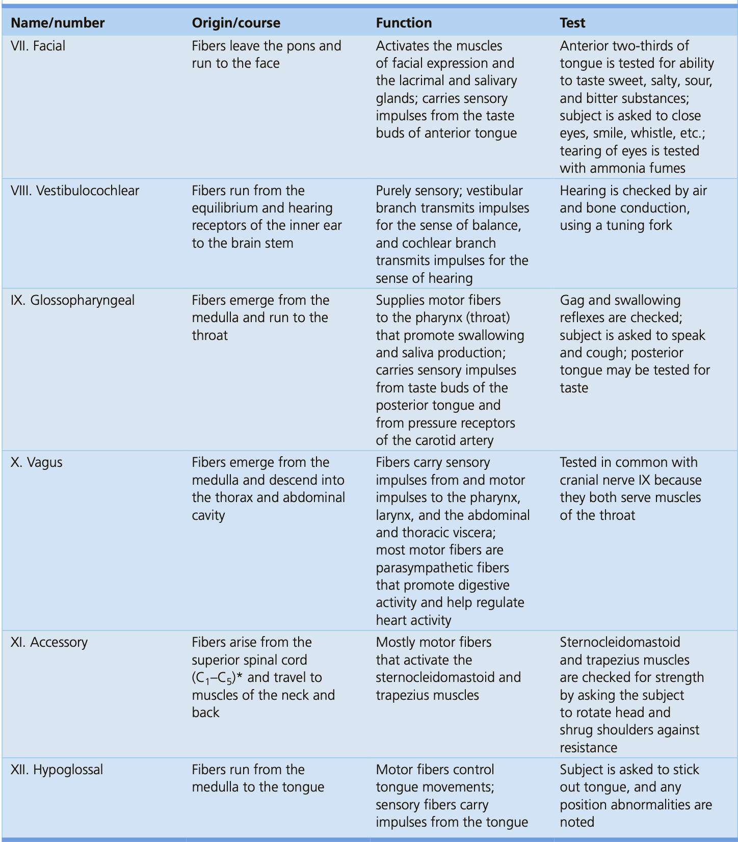
Cranial nerve 7: Facial (VII) spit cry and squint
This is the main motor nerve of the face. innervating the main muscles of facial expression, muscles of the scalp, stylohyoid muscle, platysma and stapedius muscle. The facial nerve also does taste of the anterior 2/3rds of the tongue as well as parasympathetic motor for the salivary glands and lacrimal glands (tears).
Origin/pathway
This nerve emerges from the pontomedullary justion and is between 6 and 8.
Associated Nucleus/Nuclei
The somatic motor nucleus for the facial nerve is called the facial nucleus and handles somatic motor of the face. It is located in the pons. The parasympathetic motor nucleus of the facial nerve is called the superior salivatory nerve and does control of the lacrimal glands, as well as the submandibular and sublingual salivatory glands.
This nerve also interacts with the nucleus tractus solitarus which is where it sends that 2/3rds anterior taste to.
Cranial nerve 8: Vestibulocochlear (VIII)
This is a pure sensory nerve which carries sensory information about the vestibular system as well as hearing. it is a pure sensory nerve.
Origin/pathway
This nerve emerges most laterally from the pontomedullary junction, along with Cranial nerve 6 and 7 which are medial to this nerve.
Nucleus
This nerve is pretty clead with its function and has 2 nuclei for each function. The signals for the special sense of hearing is sent to the cochlear nucleus and the signals for the (special?) sense of balance is sent to the vestibular nucleus. These nuclei are located in the lower pons to the open medulla
Cranial nerve 9: Glossopharangeal (IX)
This is a mixed information nerve, it does both motor (efferent) and sensory (afferent) information.
Sensory
This nerve is responsible for the special visceral sense of taste and general somatic sensation of the posterior 1/3rd of the tongue as well as sensory information for the upper pharynx, eustachian tube and middle ear. It also does visceral sensation for the carotids and carotid sinuses sensing chemical levels and pressure.
Motor functions
This innervates the stylopharyngeus which elevates the pharynx and larynx during swallowing. This allows it to do the gag reflex. It also allows for parasympathetic saliva production.
Origin/pathway
This originates from the post olivary sulcus, the sulcus posterior to thre olives. Nerves 9, 10, and cranial 11 all emanate from here.
Test
This is tested via gag and swallowing reflex. The subject is also asked to talk and cough. you could check taste of posterior tongue.
Associated Nucleus/Nuclei
The glossopharyngeal nerve is associated with 4 nuclei
- inferior salivatory nucleus: this parasympathetic motor association is because of its function to innervate the parotid salivary glands (lower pons)
- Nucleus tractus solitarus: this is due to it providing the special visceral sense of taste to the posterior 1/3rd of the tongue. As the glossopharangeal nerve also does carotid visceral sensation it connects here for this as well (lower pons to closed medulla)
- Spinal nucleus of V: this is due to it providing general somatic sensation to the posterior 1/3rd of tongue, upper pharynx etc (mid pons to c2)
- Nucleus ambiguus: this is a branchial motor nerve, and is a important nerve for controlling muscles of the pharynx and larynx. the association with the glossopharyngeal nerve is due to its function controlling the stylopharangeus muscle.
Cranial nerve 10: Vagus nerve (X)
The vagal nerve has a variety of functions:
Somatic sensory
It does somatic sensory, providing innervation to the skin of the external acoustic meatus (external auditory canal, or your earhole), as well as sensation to the laryngopharynx and larynx.
Special sensation
It provides the special senses of taste to the root of the tongue and the epiglottis.
Visceral sensation
it provides visceral sensory for the heart and abdominal viscera.
Somatic motor
It provides motor innervation to the majority of muscles of the pharynx soft palate and pharynx.
Parasympathetic motor
it also provides parasympathetic motor innervation ot the smooth muscle of the trachae brinchi gastro intestinal tract and regulates heart rhythm.
Test
you can test this with the glossopharyngeal test as it also innervates muscles of the throat.
Origin/pathway
This originates from the post olivary sulcus, the sulcus posterior to thre olives. Nerves 9, 10, and cranial 11 all emanate from here.
Nucleus
The Vagal nerve is associated with 4 nuclei:
- The spinal nucleus of V: This is due to its somatic sensory functions
- The nucleus tractus solitarus: At the caudal part of this nucleus both visceral sensation and taste (visceral special sensation) are processed, and thus both vagal sensory functions connect with this nucleus.
- Nucleus ambiguus: the vagus nerve controls most muscles of the pharynx and larynx and thus connects to this nucleus
- The dorsal motor nucleus of X: This is a visceral efferent nucleus which provides the visceral motor functioning for the thorax and abdomen (heart lung, GI system) This is where the well known parasympathetic functions of the vagus nerve stems from
Cranial nerve 11: Accessory nerve (XI)
This is split into 2 functions. The cranial and spinal accessory nerves. The cranial nerve originates in the nucleus ambiguus, and the spinal in the spinal accessory nucleus. the spinal portion then travels up to join with the cranial, then depart together and then once again split with the spinal innervating the trapezius and the sternomastoid muscles and (See blue section) the cranial IX travelling with the vagus and doing vagal things ( you can even think of it as an extension of the vagus.
Teaching divergence
Note: there is a divergence between john reynolds teaching of this nerve and information on the internet. john reynolds teaches that the Cr XI is essentially an extension of the vagus nerve whereas from what I can find on the web it looks to be purely branchial motor.
Cranial part XI
Origin/pathway
This originates from the post olivary sulcus, the sulcus posterior to thre olives. Nerves 9, 10, and cranial 11 all emanate from here.
Nuclei
The 3 nuclei for this nerve are:
- Nucleus ambiguus: The cranial accessory nerve definitely does branchial motor innervation of the muscles of the pharynx and larynx John reynolds says:
- The nucleus of the solitary tract: possibly associated with vagal functions
- the spinal nucleus of V: possibly associated with vagal functions
Spinal part XI
This does branchial somatic motor to the sternomastoid and trapezius muscles
Nucleus
This part of the accessory nerve attaches to the accessory spinal nucleus which is a branchial somatic efferent nucleus. This is due to its role in controlling the sternomastoid and trapezius muscles. This nucleus is found in Spinal levels C1-C5
Cranial nerve 12: Hypoglossal nerve (XII)
This innervates the tounge. Lots of muscles so has many little fibres.
Test
This is tested by getting the patient to stick their tongue out
Origin
Cranial nerve rootlets emerge from the medullary anterolateral sulcus, between the pyramids and olives
Nucleus
This nerve attaches to the general somatic efferent nucleus called the hypoglossal nucleus. it is located in the open and closed medulla