The skeletal structure of the cranium
The Skeletal structure of the cranium is somewhat simple, with 8 bones that make up the cranium. This is relates back to respiratory in MED2 and is continued more.
The cranium is the “container” which holds the brain and is made of 8 bones:
- Frontal bone
- 2x Parietal bones
- 2x Temporal bones
- Occipital bone
- Ethmoid bone
- Sphenoid bone
These are arranged as such:
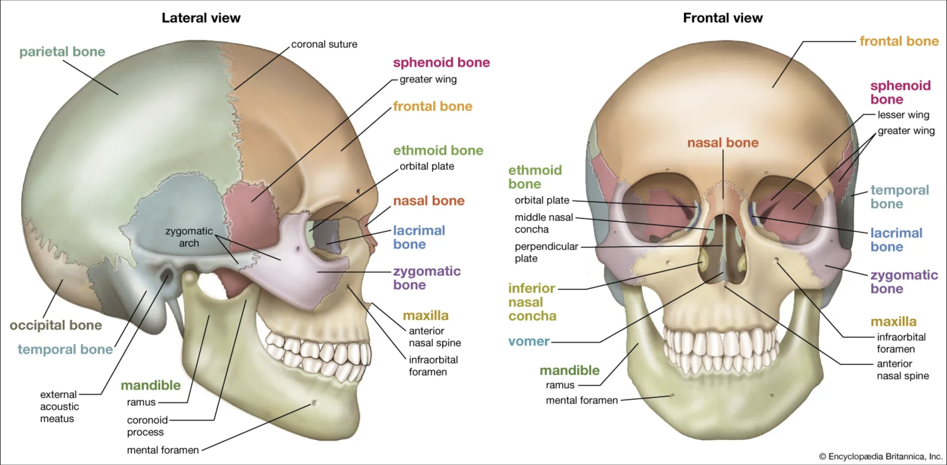
The cranial flat bones join with sutures, which are the coronal, sagittal, lamboid, and squamous. the t intersection where the coronal and sagittal suture join is called the bregma, and the t intersection where the sagittal and lambdoid sutures join is called the lambda.
In the inferior region of the internal skull there are 3 pairs of “pools” called fossa, where the frontal lobes, temporal lobes and cerebullum sit. They are easily named the anterior middle and posterior fossa. There are many structures in the medial portion of the cranial floor where there are holes for entry and exit for cranial nerves, blood vessels, sinuses and a big one for the spinal cord.
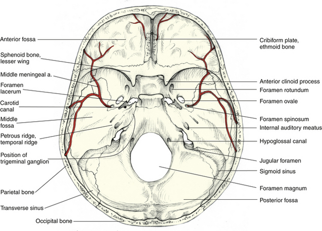 Structures of note in the cranial floor are:
- petrous bone of the temporal bone, extremely strong
- cribriform plate of the ethmoid bone, for the olfactory nerves from the olfactory bulb to pass through to the nasal cavity
- sella turcica of the sphenoid bone, for the pituitary gland to sit inside of
Foramina for the cranial nerves to pass through are listed, and are good to remember:
- Olfactory foramina (cribriform plate), this is for CNI for smell
- Optic foramen, this is for CN II for sight.
- Superior orbital fissure for CN III, IV, V (first branch), VI for oculomotor and a number of other functions. This cannot be seen on the diagram.
- Foramen rotundum for CN V (second branch)
- Foramen ovale for CN V (third branch)
- Internal auditory meatus for CN VII and VIII
- Jugular foramen this is for the CN IX X XI
- Hypoglossal canal which is for the CN XII